Explainable TI-RADS for any ultrasound.
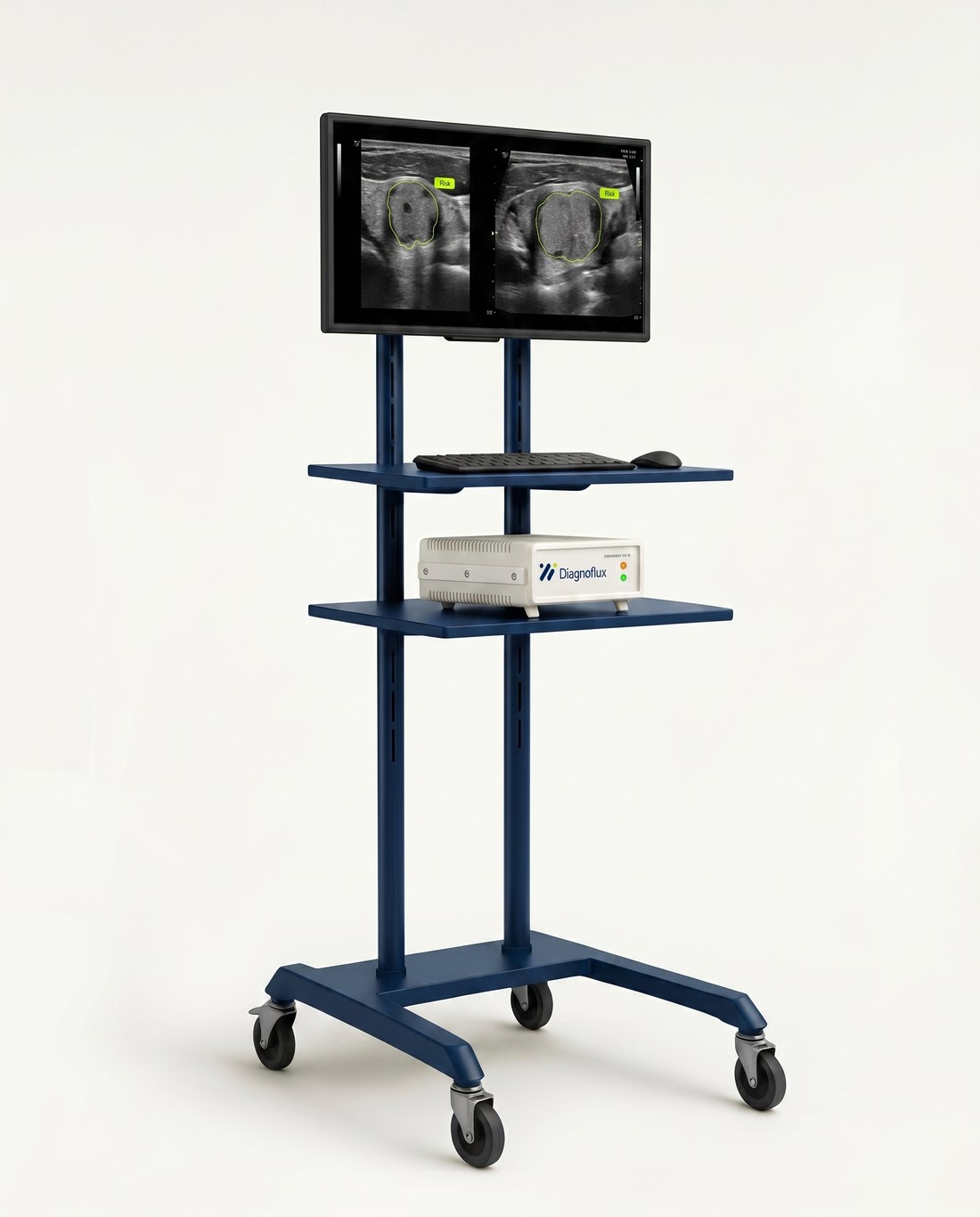
Diagnoflux reads your ultrasound's HDMI output and overlays explainable TI-RADS risk in real time. No new hardware.
Backed and validated by
Ultrasound is operator-dependent, so decisions drift.
Thyroid risk depends on who holds the probe and which device they use. The same nodule can be scored differently across readers and machines, while most real-time AI stays locked inside a few premium systems.
-
Inter-reader variability
Margin, echogenicity and composition are judged differently across readers, and by the same reader on another day.
-
Device-dependent images
Presets and dynamic range differ by machine, so the same nodule can look different from system to system.
-
Avoidable biopsies
Inconsistent thresholds drive unnecessary fine-needle biopsies and uneven follow-up between centers.
-
AI locked to premium devices
Live decision support usually ships only inside a few high-end systems. Most installed devices have none.
From an HDMI cable to explainable risk, in real time.
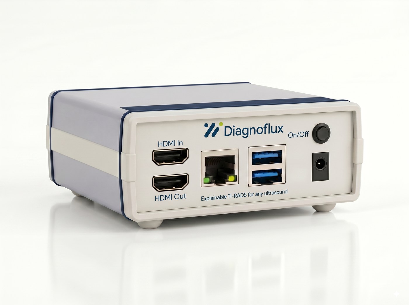
A small gateway sits between your ultrasound and a second monitor. It adds an AI layer on top of the device you already own, without touching its certified hardware or software.
-
01
Connect
Plug the gateway into your ultrasound's HDMI output and a second monitor into its HDMI out. Nothing changes on the ultrasound itself.
-
02
Analyze
Frames stream over an encrypted link to a single multi-head AI model, running in a Turkey-based cloud or on your own on-prem server.
-
03
Overlay
Segmentation and TI-RADS overlays appear on the second monitor in real time. A structured report is generated after the exam.
Explainable TI-RADS, broken down.
Diagnoflux follows the same TI-RADS workflow a clinician already uses, then shows its working at every step instead of returning a single opaque score.
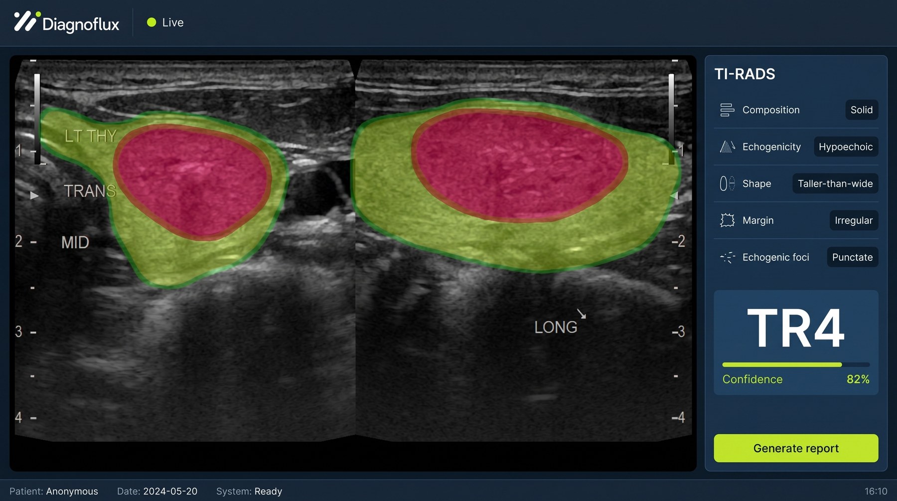
Five TI-RADS components, scored
- Composition
- Echogenicity
- Shape
- Margin
- Echogenic foci
Two planes, one TR level
You mark the representative frame in the transverse and longitudinal planes. Each component is scored and combined into the ACR TI-RADS level.
Calibrated, not a black box
Every result carries a per-component breakdown and a calibrated confidence, so you can see what drove the score rather than trusting a single number.
Reports that fit your workflow
After the exam, findings export to PDF and DICOM, ready for PACS, with the overlays and component scores attached.
Device-independent changes the math.
Because Diagnoflux sits on top of existing equipment rather than replacing it, the value shows up in cost, reach and day-to-day clinical work.
Works with the devices you own
Any ultrasound with an HDMI output joins in. No rip-and-replace, and no vendor lock-in to a single manufacturer.
Lower total cost of ownership
Upgrade through a gateway and a license instead of new capital equipment, with predictable running costs.
Fewer avoidable biopsies
Standardized, explainable reads aim to cut unnecessary fine-needle biopsies and even out follow-up between centers.
No new workflow
There is no second complex interface. Clinicians scan as usual while the overlays appear on the monitor alongside.
Your data stays where your policy needs it.
Run Diagnoflux in a Turkey-based cloud or entirely inside your own network. Either way, the link is encrypted and access is controlled and logged.
Turkey-based cloud
Managed GPUs hosted in Turkey. Frames are encrypted in transit and processed under data rules built for KVKK and GDPR.
- TLS 1.3 encrypted transport
- Short-lived JWT authentication
- Role-based access and audit logging
End-to-end latency target P95 ≤ 150 ms
On-premise
The analysis server runs inside your own network, so imaging data never leaves the building. Ideal where data residency is non-negotiable.
- Runs on your servers and network
- No data leaves your premises
- Your retention and backup policy
End-to-end latency target P95 ≤ 80 ms
Built to the right standards
Software lifecycle and risk management follow IEC 62304, ISO 13485 and ISO 14971. The software is assessed under MDR 2017/745 Rule 11, with the gateway documented as a non-diagnostic accessory.
- MDR 2017/745
- IEC 62304
- ISO 13485
- ISO 14971
- KVKK
- GDPR
Diagnoflux is an investigational device undergoing clinical validation and is not yet CE-marked. Regulatory references describe the development pathway, not a completed certification.
Built for the people in the room.
The pilot focuses on thyroid endocrinology, but the value reaches everyone who relies on the same scan, from the clinic to the quality office.
Endocrinology and radiology
Physicians performing thyroid ultrasound day to day.
Hospitals and imaging centers
Public, private and university sites, and hospital chains.
Quality and IT teams
Standardized reporting, data residency and access control.
Training programs
A shared visual reference for less-experienced readers.
One platform, beyond thyroid
The same gateway works with any HDMI optical or video system, so the approach extends well past the thyroid pilot.
- Endoscopy
- Colonoscopy
- Cystoscopy
- Bronchoscopy
- Dermatoscopy
- Breast US
- Carotid US
- Musculoskeletal US
- Liver US
Questions, answered.
The things clinical, IT and procurement teams ask first. Need something more specific? Ask the team.
No. Diagnoflux connects to your device's existing HDMI output. Nothing is installed on the ultrasound itself, so its original certification is untouched.
Your choice: a Turkey-based cloud or fully on-premise on your own servers. Both use TLS 1.3, short-lived JWT authentication, role-based access and audit logging, and are built for KVKK and GDPR.
Any ultrasound, or HDMI-output optical or video system, that exposes an HDMI output. Diagnoflux is manufacturer-independent, so mixed fleets work together.
Not yet. It is an investigational device undergoing clinical validation. The regulatory pathway follows MDR 2017/745 (Rule 11), IEC 62304, ISO 13485 and ISO 14971.
No. There is no second complex interface to learn. Clinicians scan as usual, overlays appear on a second monitor in real time, and a structured report is produced after the exam.
Each nodule gets a per-component TI-RADS breakdown (composition, echogenicity, shape, margin and echogenic foci) with calibrated confidence, mapped to the ACR TI-RADS TR1 to TR5 levels.
See Diagnoflux on your own devices.
Tell us about your setup and we will arrange a walkthrough or an on-site pilot. We typically reply within two business days.